Ophthalmology Data Annotation for AI
AI in ophthalmology is blooming with its greater potential in various aspects, including screening, diagnosis, treatment planning, and patient management. The field of ophthalmology was indeed the pioneer in adopting AI in medicine. The IDx-DR system, designed for the autonomous detection of diabetic retinopathy, was the first ever FDA-approved AI-based medical device. This is being followed by many such AI models. This being said it is important to know that no AI model could be developed without feeding in the data in the form of labels. This process of tagging or labelling is known as annotation, which is, in fact, the foundation for all AI models.
In this blog, let us brush up on the role of annotation in developing an AI model.
AI’s Role in Ophthalmology:
Screening & Diagnosis
Ophthalmology based AI systems effectively analyse and detect eye related complications. Some of them as listed as follows
-
- Diabetic Retinopathy (DR)
- Age related Muscular Degeneration (AMD)
- Glaucoma
- Cataract
- Retinal vascular Diseases
- Swollen optic disc / papilledema etc
- Ocular tumour
- Corneal disorders
Personalised Treatment Plans
Teleophthalmology (remote screening and diagnosis)
Drug Development and Research
Ophthalmology image types
The role of the dataset is crucial in developing an AI model as it directly impacts the model's performance, accuracy, and generalization capabilities. A well-chosen and properly curated dataset is the foundation upon which the AI model is built. In ophthalmology, various types of ophthalmology images are available for annotation to train and validate artificial intelligence (AI) algorithms. Some common types of images available for annotation in ophthalmology include:
Fundus Photographs:
Fundus photographs are high-resolution images of the back of the eye, capturing the retina, optic nerve, blood vessels, and other structures. These images are crucial for automatic detection of diabetic retinopathy (exudates), age-related macular degeneration, and glaucoma. A fundus photograph is typically produced using a medical imaging device called a fundus camera or retinal camera.
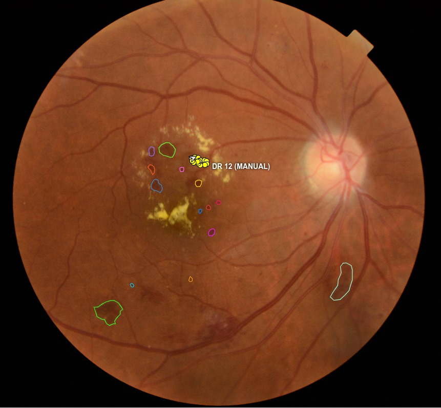
The image shows annotated diabetic retinopathy image with haemorrhages.
Optical Coherence Tomography (OCT) Images:
OCT is a non-invasive imaging technique that provides cross-sectional images of the retina, enabling a deeper understanding of the retinal structures. OCT images are used to assess retinal thickness, retinal fluids, detect macular edema, and monitor retinal disease. The device used to produce OCT images is known as an OCT machine or OCT scanner.
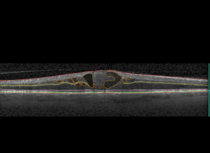
OCT image of the retinal layers and the presence of IRS fluid (marked in orange color - a sign of diabetic retinopathy).
Slit-Lamp Images:
Slit-lamp images are used to examine the anterior segment of the eye, including the cornea, iris, and lens. These images are valuable for diagnosing conditions such as corneal disorders, cataracts, iritis, and many others. Slit lamp images are produced using a medical device called a slit lamp biomicroscope or a slit lamp.
Annotation Types
When annotating ophthalmologic images, various annotation types can be used to identify and delineate specific regions or structures of interest. Some of the annotation types are as follows
- Bounding Boxes
- Semantic segmentation
- Instance segmentation
- Keypoints
- Polylines
- Landmarks
- Depthmarks
The choice of annotation type depends on the specific analysis objectives, the type of imaging data, and the complexity of the structures being annotated.
Annotation Tools
Below are some of the annotation tools which are used to annotate ophthalmology dataset.
- ITK-SNAP - Open-source software application for medical image segmentation.
- VGG Image Annotator (VIA) - Open – source image annotation tool which is customizable to suit specific annotation needs.
- 3D Slicer - 3D Slicer is a free, open-source software for visualization, processing, segmentation, registration, and analysis of medical, biomedical, and other 3D images
- Label Box - Free commercial online web-based annotation system for segmentation and classification purposes.
- Rectlabel - RectLabel is a popular image annotation tool that allows users to annotate bounding boxes and segmentation masks on ophthalmology images.
- LabelImg - Popular open-source image annotation tool that is used to create bounding boxes around the object of interest in 2d images.
- LabelMe - LabelMe is another open-source image annotation tool that allows users to draw polygons and create segmentations for objects in images
Apart from the above-mentioned tools there are other developing software platforms such as OCT – Annotation Tool which aids in automating the screening and annotation process. Ultimately, the choice of annotation tool depends on the specific requirements of the ophthalmic image dataset and the type of annotations needed.
Open Datasets available for Ophthalmology
Open datasets could be an easy and quick way to kick-start your AI model development. Some of the opensource datasets for ophthalmology are listed below:
Diabetic Retinopathy Detection : https://www.kaggle.com/c/diabetic-retinopathy-detection/data
Eye OCT Datasets : https://www.kaggle.com/datasets/kmader/eye-oct-datasets
1000 Fundus images with 39 categories : https://www.kaggle.com/datasets/linchundan/fundusimage1000/code
APTOS 2019 Blindness Detection : https://www.kaggle.com/c/aptos2019-blindness-detection/data
Haidata's experienced Ophthalmologists and Optometrists can help you deliver high quality training data for your next AI project in Opthalmology.
Write to us today at info@haidata.ai for a free sample annotation!